
![]()
Contents
What does
cardiomyopathy mean?
The various forms of
cardiomyopathy.
Heart Murmurs
& cardiomyopathy.
The Electrocardiogram &
cardiomyopathy.
The
Radiographs & cardiomyopathy.
The
Echocardiogram & cardiomyopathy.
Underlined test are linked
to a glossary, but the definition should show if you hold your mouse over the
word as well.
![]() What does cardiomyopathy mean?
What does cardiomyopathy mean?
Derivation and Definitions: cardio- = heart; myo- =
muscle; pathos- = disease
Cardiomyopathy is a
term that is used to describe diseases of the heart muscle. There are many types
of heart disease, but cats generally develop three different forms of heart
muscle disease: dilated cardiomyopathy, restrictive cardiomyopathy, and
hypertrophic cardiomyopathy. Each of these conditions is different, but
ultimately they cause problems because the heart becomes unable to pump an
adequate amount of blood to supply the body. Just like humans,
cats can have heart disease for a long time before developing heart failure. A
severe, life-threatening condition, heart failure occurs when the heart is no
longer able to pump enough blood to supply the tissues with the oxygen they
require. The right side, left side, or both sides of the heart can fail, causing
a number of complications.
One of the most severe forms of heart failure occurs when the lungs fill with
fluid, a condition called pulmonary edema. This complication occurs because the
left side of the heart is not pumping blood effectively. Excessive pressure
builds up behind the pump, and fluid leaks into the air spaces in the lungs.
Thus, the cat effectively is drowning in its own fluids, which inhibits the
exchange of oxygen between the lungs and the blood. The result is that the cells
of the body do not receive enough oxygen and begin to die. If uncorrected,
pulmonary edema leads to multiple organ failure and death.
Another complication of heart disease in cats is the development of a blood
clot, clinically known as aortic
thromboembolism, which usually forms in the
heart and travels through the blood stream. Most commonly, the clot lodges at
the branch of the aorta that feeds the back legs, shutting down blood flow and
causing partial or complete paralysis. This condition is excruciatingly painful
and requires immediate medical attention. Cats experiencing an aortic
thromboembolism will be unable to move their back legs and may vocalize due to
the pain. Aortic
thromboembolism usually indicates significant heart disease;
two thirds of cats that develop this condition will die or be put to death
humanely. In cats that survive aortic
thromboembolism, recurrence is common.
Common clinical signs include
tachypnea, panting associated with any activity,
dyspnea, coughing, anorexia, vomiting, weight loss and lethargy. Some cats will
develop hind leg paralysis, loss of femoral pulses, and cool limbs due to
thromboembolism. Syncope or sudden death may also occur. Often, a heart murmur,
gallop rhythm, or abnormal lung sounds are detected on
auscultation of the
heart. The cat may have experienced recent stresses such as anesthesia, surgery,
boarding, or car rides that caused it to develop heart failure.
![]()
Click on the heart to go back to the top of the page.
![]() The various forms of
cardiomyopathy:
The various forms of
cardiomyopathy:
A primary (idiopathic) cardiomyopathy is a disease of heart muscle with no known
underlying
etiology. Primary Cardiomyopathies are diagnosed by the
morphologic and functional
appearance of the patient’s heart and ruling out other causes for these
“patterns”.
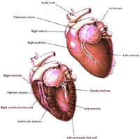
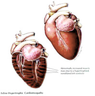
Normal heart
HCM heart
- Hypertrophic cardiomyopathy (HCM):
concentric (symmetric or asymmetric)
hypertrophy.
Hypertrophic
cardiomyopathy, the most common form of feline heart muscle disease, occurs
when the lower left chamber of the
heart, called the left ventricle, thickens and stiffens, while
the top left chamber, the left
atrium, enlarges. This thickened left ventricle does not leave much
room in the chamber to fill with
blood. Thus, smaller than normal amounts of blood are
pumped out of the heart with each
contraction. Additionally, this thickening of the heart muscle
increases the heart's own consumption
of oxygen, which is needed to supply the additional
muscle present. If these oxygen
demands are not met, then cell death occurs and leads to
areas of scarring in the heart
muscle.
Normal
heart
DCM heart
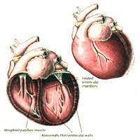
- Idiopathic Dilated cardiomyopathy (DCM): eccentric hypertrophy with
myocardial failure.
Dilated
cardiomyopathy, occurs when the heart chambers become big and dilated, like a
"flabby balloon." With dilation, the
heart muscle is often weakened dramatically so that it
cannot contract with the normal
amount of force. This disease was very common before it
was recognized that the majority of
cases were due to a dietary deficiency of the amino acid
taurine. Since commercial diets now
adequately are supplemented with taurine, this disease
is uncommon.
- Restrictive cardiomyopathy (RCM).
Restrictive cardiomyopathy has also been called intermediate cardiomyopathy
because it has
characteristics of both dilated and
hypertrophic cardiomyopathy. With this form of the illness,
the walls of the cat's heart develop
fibrosis, which is the replacement of normal heart tissue
with scar tissue that does not
function as well. This scarring makes the heart stiff and less
effective as a pump.
A secondary cardiomyopathy is a heart muscle disease resulting from other
disease processes. Proven or strongly suspected causes of
Secondary Cardiomyopathy in Cats:
- Nutritional (taurine deficiency).
- Metabolic (hyperthyroidism,
acromegaly).
- Infiltrative (neoplasia,
amyloidosis).
- Inflammatory (toxins, immune reactions, infectious agents).
- Genetic (strong evidence in HCM in some breeds; may play a
role in
the susceptibility to taurine deficiency induced myocardial
failure).
- Toxic (doxorubicin, heavy metals).
Within each class, wide ranges of
morphologic and clinical presentations are
seen. The lines between forms and classifications begin to blur as the facts are
unraveled.
![]() Click on the heart to go back to the top of the page.
Click on the heart to go back to the top of the page.
 Heart Murmurs and cardiomyopathy:
At the very least,
breeding cats should be examined by a vet with a stethoscope for heart murmurs
or arrhythmias once yearly. Any cat with an abnormality should have an
echocardiogram.
A significant percentage of cats with HCM will not have a
heart murmur, however.
Grade 1
A very soft murmur
only detected after very careful auscultation
Grade 2
A soft murmur that
is readily evident
Grade 3
A moderately intense
murmur not associated with a palpable precordial thrill (vibration)
Grade 4
loud murmur; a
palpable precordial thrill is not present or is intermittent
Grade 5
A loud cardiac
murmur associated with a palpable precordial thrill; the murmur is not audible
when the stethoscope is lifted from the thoracic body wall
Grade 6
A loud cardiac
murmur associated with a palpable precordial thrill and audible even when the
stethoscope is lifted from the thoracic wall
![]()
Click on the heart to go back to the top of the page.
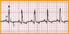 The Electrocardiogram:
Indications for electrocardiography
include arrhythmias heard on auscultation, breathing problems, shock, fainting
or seizures, cardiac murmurs, and systemic disease that affects the heart (e.g.
tumors, kidney dysfunction, heartworm disease). Electrocardiography is also
useful as part of the preoperative work-up in older animals, for monitoring
patients during and after surgery, and for evaluating the effects of cardiac
drugs. An electrocardiogram (ECG) is the only test that can
accurately diagnose an arrhythmia or a conduction abnormality. And an ECG will
help you decide when other diagnostic tests should be done, including, thoracic
radiography, or even echocardiography.
![]() Click on the heart to go back to the top of the page.
Click on the heart to go back to the top of the page.


Click on the box to the left for a link to the article.
The article show
both x-rays and echocardiograms.

The
Radiographs:
Chest
radiographs (x-rays) of asymptomatic cats may appear normal or may show mild
enlargement of the heart. In cats with clinical signs of HCM there may be
greater enlargement of the heart as well as evidence of fluid buildup in the
lungs and chest cavity.
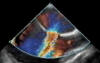
The
Echocardiogram:
This is the most
important diagnostic tool the vets have for HCM. An echocardiogram is a test in
which ultrasound is used to examine the heart. In
addition to providing single-dimension images, known as M-mode echo that allows
accurate measurement of the heart chambers, the echocardiogram also offers far
more sophisticated and advanced imaging. This is known as two- dimensional (2-D)
Echo and is capable of displaying a cross-sectional "slice" of the beating
heart,
including the chambers, valves and the major blood vessels that exit from the
left and right ventricle. Doppler is a special part of the ultrasound
examination that assess blood flow (direction and velocity). In contrast, the
M-mode and 2-D Echo evaluates the size, thickness and movement of heart
structures (chambers, valves, etc.). During the Doppler examination, the
ultrasound beams will evaluate the flow of blood as it makes it way though and
out of the heart. This information is presented visually on the monitor (as
color images or grayscale tracings) and also
as a series of audible signals with a swishing or pulsating sound.
![]() Click on the heart to go back to the top of the page.
Click on the heart to go back to the top of the page.
![]()
![]()
![]()
![]()
![]()